

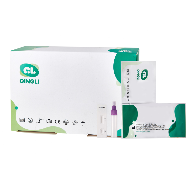
T-Ferritin Rapid Test Device (Feces)
INTRODUCTION
1,2 Tf is a type of β1 globulin, with a molecular weight of 77 KD, accounts for about 0.3% to 0.5% of the total plasma protein. It is mainly synthesized in liver, and the main function of the protein is to transport extracellular iron into cells through membrane receptor-mediated endocytosis.3 As in the case of Transferritin, serum Tf may be leaked into gastrointestinal tract during the course of bleeding. However, compared with Transferritin, Tf is more stable .4, 5 The TF Transferritin Rapid Test Device(Feces)is a rapid test to qualitatively detect low levels of Transferritin of fecal occult blood in feces. The test uses double antibody sandwich assay to selectively detect as low as 25ng/mL Transferritin of human occult blood in feces. In addition, unlike the guaiac assays, the accuracy of the test is not affected by the diet of the patients.
PRINCIPLE
-
The Transferritin Rapid Test Device(Feces) is a qualitative, lateral flow immunoassay for the detection of human occult blood in feces. The membrane is pre-coated with anti-Transferritin antibody on the test line region of the strip. During testing, the specimen reacts with the particle coated with anti-Transferritin antibody. The mixture migrates upward on the membrane chromatographically by capillary action to react with anti-Transferritin antibody on the membrane and generate a colored line. The presence of this colored line in the test region indicates a positive result, while its absence indicates a negative result. To serve as a procedural control, a colored line will always appear in the control line region indicating that the proper volume of specimen has been added and membrane wicking has occurred.
DIRECTIONS FOR USE
1)Use the specimen collection container for specimen collection. Best results will be obtained if the assay is performed within 4 hours after collection.
2)Unscrew and remove the dilution tube applicator. Be careful not to spill or spatter solution from the tube. Collect specimens by inserting the applicator stick into at least 3 different sites of the feces to collect approximately 50 mg of feces (equivalent to 1/4 of a pea).
3)Place the applicator back into the tube and screw the cap tightly. Be careful not to break the tip of the dilution tube.
4)Shake the specimen collection tube vigorously to mix the specimen and the extraction buffer.
Specimens prepared in the specimen collection tube should be tested in 1 hour after preparation.
1)Remove the test from its sealed pouch, and place it on a clean, level surface.
2)Using a piece of tissue paper, break the tip of the dilution tube. Hold the tube vertically and dispense 2-3 drops of solution (about 80 μL to 120 μL) into the specimen well (S) of the test device. Avoid trapping air bubbles in the specimen well (S), and do not drop any solution in observation window.
As the test begins to work, you will see color move across the membrane.
3)Wait for the colored band(s) to appear. The result should be read at 5 minutes. Do not interpret
the result after 10 minutes.